What is digital Imaging
Digital imaging allows us to take very detailed images of your teeth and jaws. Our dentists use these images to examine, detect, monitor, diagnose and treat any issues that are not immediately visible to the naked eye.
Perfect imaging on-site
or call (07) 3266 7120
Advantages of digital imaging
Unlike traditional X-rays, which had to be developed and displayed on special plates, digital images are electronically transferred directly onto the dentist’s computer. This allows them to be viewed and analysed in minutes rather than having to wait for days to get images back from the lab.
Digital transfer of the images means that they can easily be manipulated to improve contrast and give a more detailed picture of your teeth and periodontal areas.
Plus, our dentists can easily retrieve images right beside you while you’re in the chair. This makes it easy to point out any concerns and allows us to explore your options together.
Electronic storage also allows dentists to easily retrieve previously taken images for comparison.
And, since digital images require none of the processing chemicals and hazardous materials disposal required by traditional images, they are far more eco-friendly.
What digital imaging means for patients is that our dentists can take immediate preventative and curative action early on. This allows us to strengthen and preserve teeth so you’re able to enjoy a healthy natural smile and dental function for years to come.


Not just X-rays
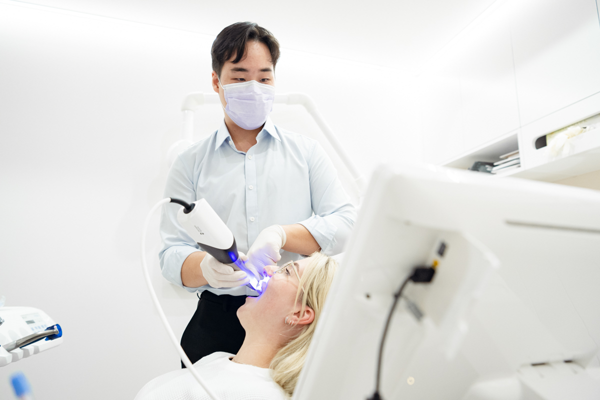
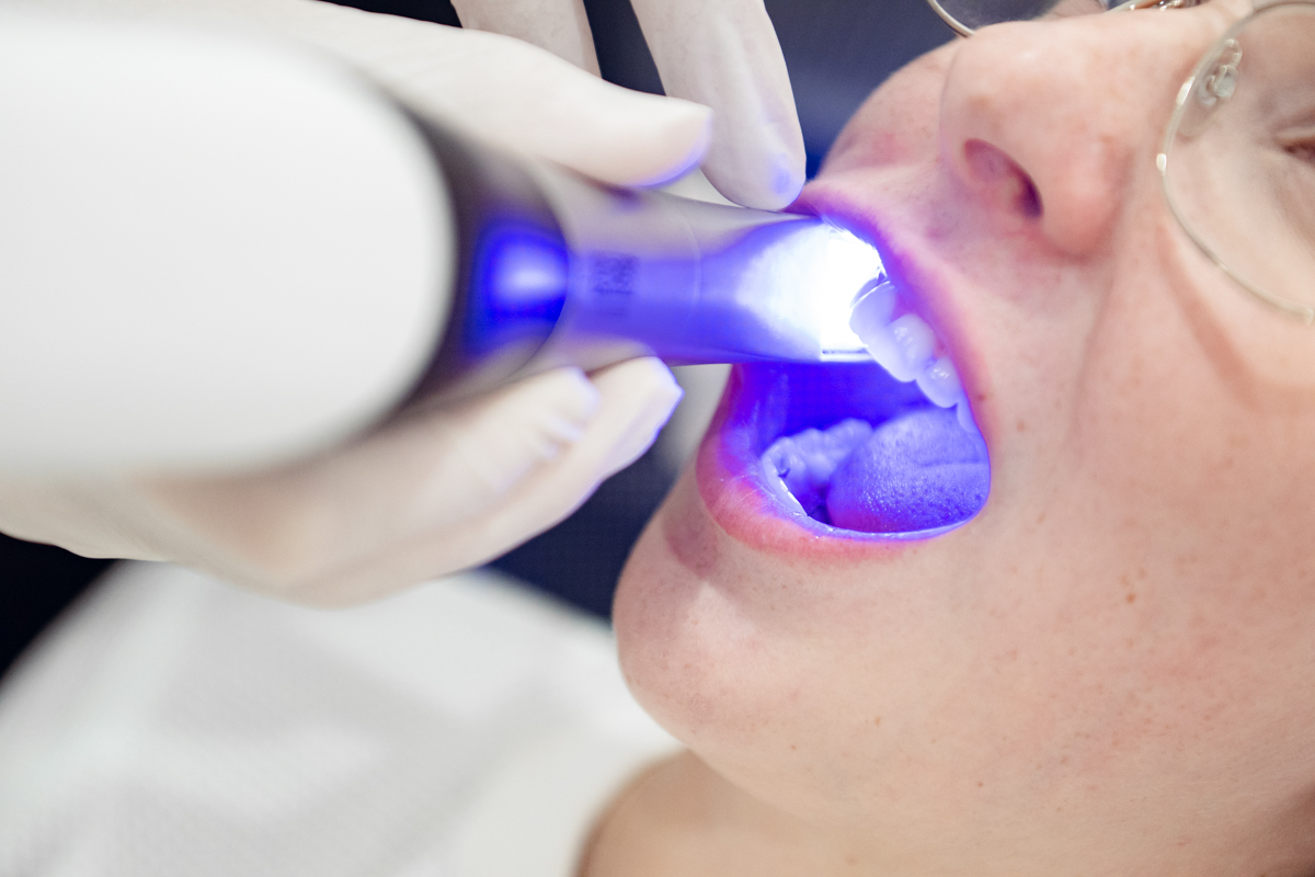
Ivy Dental uses digital scanners to create a 3D digital model of your teeth. This means that for many cases, we can avoid taking an impression, instead capturing images of the teeth and gums using a handheld wand.
The scanner uses light to capture images of the teeth and gums, and the images are instantly displayed on a computer screen. The images captured by the scanner are processed by specialised software that creates a 3D digital model. The software stitches together the individual images captured by the scanner to create a complete and accurate representation of what we’re trying to capture.
The digital model can be viewed and manipulated on the computer screen, allowing your dentist to make any necessary adjustments. Your dentist can also use the digital model to show you what your teeth will look like after your treatment is completed.
From here, our lab technicians take over, using these 3D scans, in combination with photos and videos to create the perfect smile for your face. Paying particular attention to the contours of your lips and gums, shape of your existing teeth and the facial structures, we can create the most flattering smile that is bespoke for you.
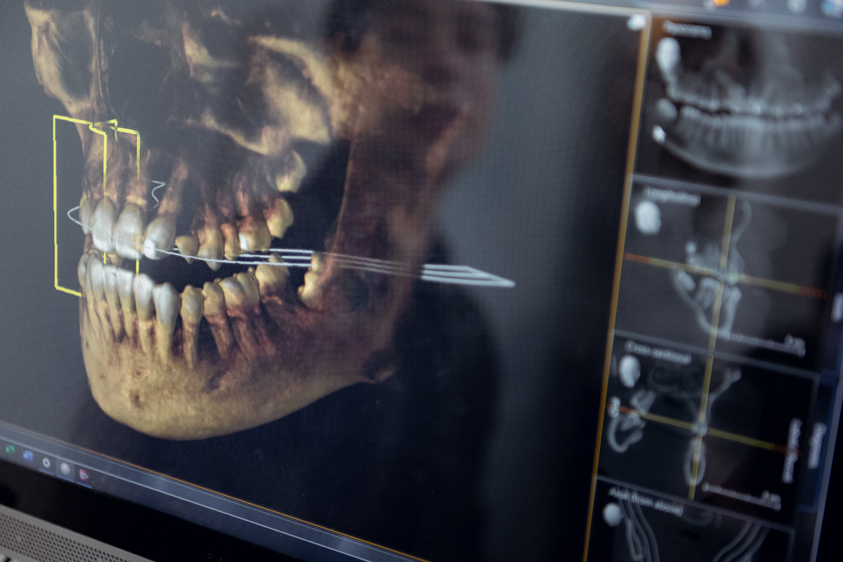
3D Imaging
CBCT goes beyond regular X-ray imaging to provide us with 3D images of your teeth, soft tissues, nerve pathways and bones in a single scan.
Especially useful for planning treatment in more complex cases, images obtained using CBCT allow us to create a more precise treatment plan tailored to your specific requirements.
Using these radiographs allows our dentists to have better outcomes when it comes to implant placement and oral surgery, orthodontics, endodontics periodontics and to diagnose and treat a range of issues, from toothaches to TMD.
LATCEPH images are taken of the sides of the face in very precise positions. They allow dentists to take specific measurements which help them to determine the current and future relationships between teeth in the top and bottom jaws. This alignment affects the patient’s bite and is useful in assessing the need for and planning orthodontic treatments.


Get in touch
Got a dental emergency?
Call us on (07) 3266 7120.
Ready to book your appointment? Head over to our online booking page.
Got questions about our services? Please fill in the form below and we’ll get right back to you.


