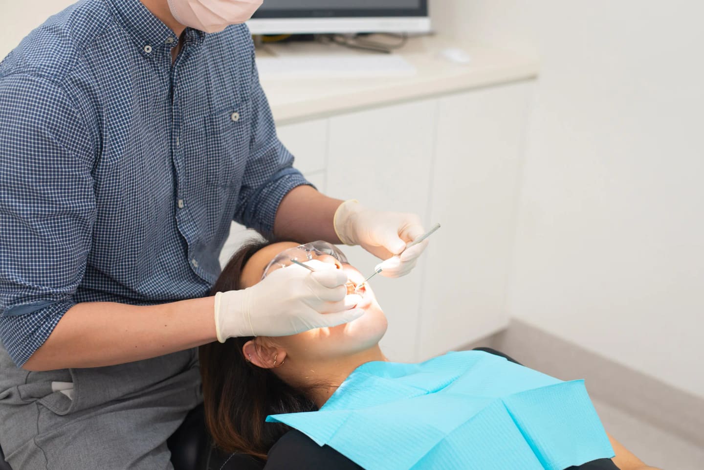
Dental X-rays are one of the most common questions patients raise, particularly parents and people who have not visited the dentist in a while. The concern usually centres on radiation exposure. It is a fair question, and the answer is straightforward.
This guide covers the types of X-rays used in dental practice, how much radiation they involve, when they are needed, and the specific considerations for pregnancy and children.
Why dentists need X-rays
A clinical examination tells your dentist a lot, but it cannot show everything. X-rays reveal problems that are invisible to the naked eye during a standard check-up:
- Decay between teeth (the most common use)
- Bone loss around teeth from gum disease
- Infections or abscesses at the root tips
- The position of unerupted teeth, including wisdom teeth
- Cysts, tumours, or other pathology in the jaw
- The condition of existing fillings, crowns, and root canal treatments
- Bone volume and density for dental implant planning
Without X-rays, your dentist would be making treatment decisions with incomplete information. Many problems are only detectable on imaging once they reach a certain size, and catching them earlier generally means simpler, less invasive treatment.
Types of dental X-rays
Bitewing X-rays
The most common type. These show the upper and lower back teeth in a single image. They are primarily used to detect decay between teeth and to check the fit of existing restorations. A typical set involves two or four images.
Radiation dose: Approximately 0.005 mSv per image with digital equipment.
Periapical X-rays
These show the full length of a tooth, from the crown to the root tip and the surrounding bone. They are used to assess root health, detect abscesses, and evaluate bone levels around individual teeth.
Radiation dose: Approximately 0.005 mSv per image.
Panoramic X-ray (OPG)
A single wide image that shows all teeth, both jaws, the jaw joints (TMJ), and the sinuses. An OPG is useful for an overall assessment, particularly for new patients, orthodontic planning, wisdom tooth evaluation, and implant assessment.
Radiation dose: Approximately 0.01 to 0.03 mSv.
Cone Beam CT (CBCT)
A three-dimensional scan that provides detailed cross-sectional images of the teeth, bone, and surrounding structures. CBCT is used for complex treatment planning, including implant placement, difficult extractions, and assessment of jaw pathology. It is not used routinely and is only recommended when 2D imaging does not provide sufficient information.
Radiation dose: Approximately 0.03 to 0.20 mSv depending on the field of view and machine settings.
Putting radiation levels in perspective
Radiation from dental X-rays is measured in millisieverts (mSv). To put dental X-ray doses in context:
- Annual background radiation in Australia: Approximately 1.5 mSv. This comes from natural sources including cosmic radiation, radon gas, and naturally occurring radioactive elements in soil and food.
- A set of 4 bitewing X-rays: Approximately 0.02 mSv. Equivalent to roughly a few hours of natural background exposure.
- One OPG: Approximately 0.01 to 0.03 mSv.
- A domestic flight (Sydney to Brisbane): Approximately 0.01 mSv from cosmic radiation at altitude.
A full set of dental X-rays delivers a fraction of the radiation you receive from your natural environment over the course of a year. Digital X-ray equipment, which is used at Ivy Dental, reduces radiation doses by up to 80% compared to traditional film-based systems.
Digital vs film X-rays
Most modern dental practices in Brisbane, including Ivy Dental, use digital X-ray sensors rather than traditional film.
The advantages of digital imaging include:
- Lower radiation. Digital sensors are more sensitive than film, so less radiation is needed to produce a diagnostic image.
- Instant results. Images appear on screen within seconds. No darkroom processing required.
- Enhanced viewing. Images can be magnified, adjusted for contrast, and compared side by side with previous X-rays.
- Easy sharing. Digital images can be sent to specialists or other providers electronically.
- No chemical waste. Film processing involves chemical solutions that require careful disposal.
At your first visit to Ivy Dental, your X-rays will appear on the screen beside you so your dentist can walk you through the findings in real time.
How often are X-rays needed?
There is no single answer. Your dentist recommends X-ray frequency based on your individual risk profile.
Typical guidelines for adults:
- Bitewing X-rays: Every 12 to 24 months for patients with moderate decay risk. Patients with low decay risk and healthy gums may extend to 24 to 36 months.
- OPG: Every 3 to 5 years for a comprehensive overview, or as needed for specific diagnostic purposes.
- Periapical X-rays: As needed based on symptoms or findings.
- CBCT: Only when clinically indicated for treatment planning.
For children and adolescents: X-ray frequency may be higher because teeth are still developing and children tend to have a higher rate of new decay. See our kids dentistry page for more on children's dental care. Your dentist will balance the diagnostic benefit against the goal of minimising radiation exposure.
Your dentist should explain why a particular X-ray is recommended before taking it. If you are unsure, ask.
Pregnancy and dental X-rays
The radiation dose from dental X-rays is very low, and the Australian Dental Association advises that dental imaging is generally considered safe during pregnancy when clinically necessary. Modern digital equipment and lead apron shielding further minimise exposure.
That said, most dentists prefer to take a conservative approach:
- Routine X-rays are usually deferred until after delivery when possible.
- If X-rays are needed for diagnosis during pregnancy (for example, to assess an infection or emergency), they can typically be taken safely with appropriate shielding.
- The second trimester is generally preferred for any necessary dental treatment.
If you are pregnant or think you may be, let your dentist know at the start of your appointment. They will adjust the treatment plan accordingly.
Safety measures at the practice
Dental practices follow strict protocols to minimise radiation exposure:
- Lead apron and thyroid collar. These shield the body from scatter radiation during imaging.
- Rectangular collimation. Modern X-ray heads restrict the beam to the area being imaged, reducing unnecessary exposure.
- Digital sensors. Require significantly less radiation than film.
- Justification principle. X-rays are only taken when there is a clinical reason. Your dentist should be able to explain why each image is needed.
- ALARA principle. Radiation exposure is kept As Low As Reasonably Achievable. This is a standard applied across all medical and dental imaging in Australia.
When to ask questions
You are entitled to ask why an X-ray is being recommended and what it will show. A good dentist will explain the diagnostic purpose clearly. You can also ask:
- When were my last X-rays taken?
- What are you looking for with this image?
- Can we use my existing X-rays from another practice?
At Ivy Dental, we are happy to discuss imaging decisions with you. We are open seven days at our Everton Park and Nundah practices in north Brisbane, with our general dentistry team available at these clinics.
Book a consultation or call (07) 3266 7120.